Functional microscopic anatomy of the kidney and bladder is a captivating field that delves into the intricate structural and functional aspects of these vital organs. This exploration unveils the histological architecture of the renal corpuscle, proximal convoluted tubule, loop of Henle, and distal convoluted tubule, shedding light on their roles in maintaining water balance and filtering waste products.
Simultaneously, the bladder’s histological structure, including the urothelium, lamina propria, and muscularis, is examined, highlighting its role in urine storage and expulsion.
By understanding the functional microscopic anatomy of the kidney and bladder, medical professionals gain invaluable insights into the diagnosis and treatment of various diseases. Imaging techniques, such as ultrasound and MRI, play a crucial role in visualizing these organs, aiding in the detection and monitoring of abnormalities.
Functional Microscopic Anatomy of the Kidney

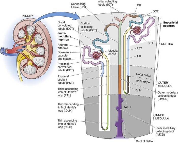
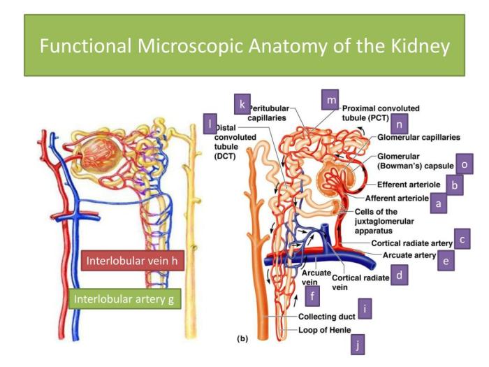
The kidney is responsible for filtering blood and producing urine. It is composed of millions of microscopic functional units called nephrons. Each nephron consists of a renal corpuscle and a renal tubule.
Renal Corpuscle
The renal corpuscle is the site of filtration. It consists of a glomerulus, which is a tuft of capillaries, and Bowman’s capsule, which is a cup-shaped structure that surrounds the glomerulus. The glomerular capillaries are lined by fenestrated endothelium, which allows blood to be filtered into Bowman’s capsule.
Proximal Convoluted Tubule, Functional microscopic anatomy of the kidney and bladder
The proximal convoluted tubule (PCT) is the first part of the renal tubule. It is responsible for reabsorbing most of the water, sodium, and glucose from the filtrate. The PCT also secretes hydrogen ions and other substances into the filtrate.
Loop of Henle
The loop of Henle is a U-shaped structure that descends deep into the medulla of the kidney and then ascends back to the cortex. The descending limb of the loop of Henle is permeable to water, while the ascending limb is impermeable to water.
This creates a concentration gradient in the medulla, which allows the kidney to concentrate urine.
Distal Convoluted Tubule and Collecting Ducts
The distal convoluted tubule (DCT) is the last part of the renal tubule. It is responsible for fine-tuning the composition of the urine. The DCT also secretes potassium ions into the filtrate. The collecting ducts collect urine from the DCTs and transport it to the renal pelvis.
Functional Microscopic Anatomy of the Bladder

The bladder is a hollow organ that stores urine. It is lined by a layer of urothelium, which is a transitional epithelium that can stretch to accommodate changes in bladder volume. The lamina propria is a layer of connective tissue that lies beneath the urothelium.
The muscularis is a layer of smooth muscle that surrounds the bladder.
Trigone
The trigone is a triangular area at the base of the bladder. It is the site of the ureteral orifices, which are the openings of the ureters into the bladder. The trigone also contains the internal urethral orifice, which is the opening of the urethra into the bladder.
Innervation
The bladder is innervated by the pelvic nerves. The pelvic nerves control the contraction and relaxation of the bladder muscles.
Comparison of the Kidney and Bladder

| Structure | Kidney | Bladder ||—|—|—|| Function | Filter blood and produce urine | Store urine || Lining | Urothelium | Urothelium || Lamina propria | Connective tissue | Connective tissue || Muscularis | Smooth muscle | Smooth muscle || Trigone | Yes | Yes || Ureteral orifices | Yes | No || Internal urethral orifice | No | Yes |
Clinical Applications
Understanding the functional microscopic anatomy of the kidney and bladder can aid in diagnosing and treating diseases. For example, knowing the structure of the renal corpuscle can help doctors diagnose glomerulonephritis, which is a disease that affects the glomeruli. Knowing the structure of the bladder can help doctors diagnose cystitis, which is a disease that affects the bladder.Imaging
techniques can be used to visualize the kidney and bladder. For example, ultrasound can be used to visualize the kidneys and bladder in real time. Computed tomography (CT) scans and magnetic resonance imaging (MRI) scans can be used to create detailed images of the kidneys and bladder.
Query Resolution: Functional Microscopic Anatomy Of The Kidney And Bladder
What is the functional unit of the kidney?
The nephron is the functional unit of the kidney, responsible for filtering blood and producing urine.
What is the role of the loop of Henle in the kidney?
The loop of Henle plays a crucial role in maintaining water balance by creating a concentration gradient in the kidney medulla.
What is the histological structure of the bladder wall?
The bladder wall consists of three layers: the urothelium, lamina propria, and muscularis.